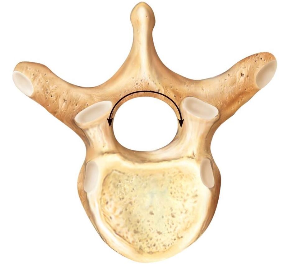
Together with the increasing convexity of the posterior aspect of the vertebral body, these changes in the position of the pedicles alter the shape of the normal bony spinal canal from an ellipse at L1 to a triangle at 元 and more or less a trefoil at L5 (Fig. This narrows the anteroposterior diameter and widens the transverse diameter of the vertebral canal from above downwards. From L1 to L5, the pedicles become shorter and broader, and are more lateral. Together with the broad and flat lamina, they form the vertebral arch. The two pedicles originate posteriorly and attach to the cranial half of the body. Meningeal branches of spinal nerves innervate all vertebrae Pedicles A sagittal cut through the vertebral body shows the endplates to be slightly concave, which consequently gives the disc a convex form. This is the cartilaginous endplate, forming the transition between the cortical bone and the rest of the intervertebral disc. A layer of cartilage covers this central zone, which is limited by the peripheral ring.

The central zone – the bony endplate – shows many perforations, through which blood vessels can reach the disc. At the upper and lower surfaces, two distinct areas can be seen: each is a peripheral ring of compact bone – surrounding and slightly above the level of the flat and rough central zone – which originates from the apophysis and fuses with the vertebral body at the age of about 16. From L1 to L5, the posterior aspect changes from slightly concave to slightly convex, and the diameter of the cylinder increases gradually because of the increasing loads each body has to carry. The curvature of articular facets is thought to assist in the stabilization and weight-bearing capacity of lumbar vertebrae.Įach vertebral body is more or less a cylinder with a thin cortical shell, which surrounds cancellous bone.The mammillary processes provide a point of attachment for intertransversarii muscles and multifidus.


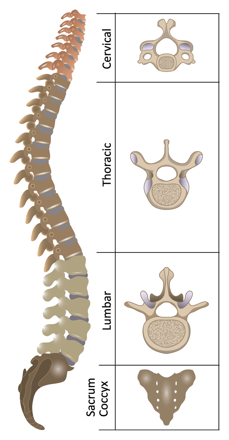
Addition of the mammillary process on the posterior aspect of the superior articular process.This is one feature that differentiates lumbar vertebrae to from thoracic Facets also have the unique feature of a curved articular surface.Articular facets are markedly vertical, with the superior facets directed posteromedially and medially.Spinous process is short and thick, relative to the size of the vertebra, and projects perpendicularly from the body.Typical lumbar vertebrae have several features distinct from those typical of cervical or thoracic vertebrae. It also has a complicated innervation and vascular supply. The complex anatomy of the lumbar region is a remarkable combination of these strong vertebrae (with their multiple bony elements) linked by joint capsules, and flexible ligaments/tendons, large muscles, and highly sensitive nerves.The intervertebral discs, along with the laminae, pedicles and articular processes of adjacent vertebrae, create a space through which spinal nerves exit. The intervertebral discs are responsible for the mobility without sacrificing the supportive strength of the vertebral column.The lumbar vertebrae, as a group, produce a lordotic curve The lumbar spine consists of 5 moveable vertebrae (numbered L1-L5). The spine extends from the skull to the coccyx and includes the cervical, thoracic, lumbar, and sacral regions.Thirty-one pairs of nerves are rooted to the spinal cord and they control body movements and transmit signals from the body to the brain. Ligaments hold the vertebrae in place, and tendons attach the muscles to the spinal column. The spaces between the vertebrae are maintained by intervertebral discs that act like shock absorbers throughout the spinal column to cushion the bones as the body moves.

The lower back (where most back pain occurs) includes the five vertebrae in the lumbar region and supports much of the weight of the upper body.


 0 kommentar(er)
